Working in the Community to Reduce Health Disparities

Neighbouring cells help cancers dodge drugs

Cancers can resist destruction by drugs with the help of proteins recruited from surrounding tissues, find two studies published by Naturetoday. The presence of these cancer-assisting proteins in the stromal tissue that surrounds solid tumours could help to explain why targeted drug therapies rapidly lose their potency.

Targeted cancer therapies are a class of drugs tailored to a cancer’s genetic make-up. They work by identifying mutations that accelerate the growth of cancer cells and selectively blocking copies of the mutated proteins. Although such treatments avoid the side effects associated with conventional chemotherapy, their effectiveness tends to be short-lived. For example, patients treated with the recently approved drug vemurafenib initially show dramatic recovery from advanced melanoma, but in most cases the cancer returns within a few months.

Many forms of cancer are rising in prevalence: for example, in the United States, the incidence of invasive cutaneous melanoma — the deadliest form of skin cancer — increased by 50% in Caucasian women under 39 between 1980 and 2004. So there is a pressing need to work out how to extend the effects of targeted drug therapies. But, until now, researchers have focused on finding the mechanism of drug resistance within the cancerous cells themselves.

Two teams, led by Jeff Settleman of Genentech in South San Francisco, California, and Todd Golub at the Broad Institute in Cambridge, Massachusetts, expanded this search into tumours’ surrounding cellular environment.

Settleman’s team tested 41 human cancer cell lines, ranging from breast to lung to skin cancers. The researchers found that 37 of these became desensitized to a handful of targeted drugs when in the presence of proteins that are usually found in the cancer’s stroma, the supportive tissue that surrounds tumours. In the absence of these proteins, the drugs worked well1. By growing cancer cells along with cells typically found in a tumour’s immediate vicinity, Golub and his colleagues showed that these neighbouring cells are the likely source of the tumour-aiding proteins2.

Protein culprit

One of the most startling results of the teams’ experiments was the discovery that a protein called hepatocyte growth factor (HGF) boosts melanoma’s resistance to treatment with vemurafenib. Intrigued by this result, both teams looked at blood samples from people who had undergone treatment with vemurafenib, and found the higher a patient’s HGF levels, the less likely they were to remain in remission.

Martin McMahon, a cancer biologist at the University of California, San Francisco, who was not affiliated with either study, explains that the results have immediate implications for the design of clinical trials, which he says could combine targeted drug therapy with drugs capable of knocking down the production of proteins such as HGF.

“These papers show that the influence of the cell’s microenvironment is important not only for melanoma, but also for pancreatic, lung and breast cancer,” McMahon says, adding that they are “very exciting, because they expand the focus of where we should be looking for the mechanisms of drug resistance”.

:: Read original here ::

Vaccine developed against Ebola

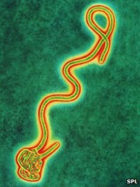
Scientists have developed a vaccine that protects mice against a deadly form of the Ebola virus.

First identified in 1976, Ebola fever kills a majority of the people it infects.

The researchers say that this is the first Ebola vaccine to remain viable long-term and can therefore be successfully stockpiled.

The results are reported in the journal Proceedings of National Academy of Sciences.

Ebola is transmitted via bodily fluids, and can become airborne. Sufferers experience nausea, vomiting, internal bleeding and organ failure before they die.

Although few people contract Ebola each year, its effects are so swift and devastating that it is often feared that it could be used against humans in an act of terroism.

All previously developed vaccines have relied on injecting intact, but crippled, viral particles into the body.

Long-term storage tends to damage the virus, paralysing the vaccine’s effectiveness.

The new vaccine contains a synthetic viral protein, which prompts the immune system to better recognise the Ebola virus, and is much more stable when stored long-term.

The vaccine protects 80% of the mice injected with the deadly strain, and survives being “dried down and frozen,” said biotechnologist Charles Arntzen from Arizona State University who was involved in its development.

He said the next step is to try the vaccine on a strain of Ebola that is closer to the one that infects humans.

:: Read original here ::

Researchers switch on genes with blue pulse

Scientists have developed a technique that could be used to deliver precise doses of hormones to people who don’t make them naturally.

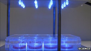
To do this, they rewired kidney cells with light-sensitive molecules from the eye, they reported in the journal Science.

When pulsed with blue light, these cells churned out proteins on demand.

Ultimately, this technique could avoid the need for people with diabetes to inject themselves regularly.

“When I speak to diabetes patients they say that if you could take away always having to inject themselves it would really increase their quality of life,” said lead author Martin Fussenegger, a bioengineer of the Swiss Federal Institute of Technology, Zurich.

Dr Fussenegger thought he saw a solution in his own field of optogenetics. Optogenetics, as the name suggests, uses light to control the behaviour cells.

To get a cell to respond to light you first have to rejigger it so it has a light-sensitive molecule on its surface. Dr Fussenegger coaxed kidney cells to express melanopsin, a molecule usually found in animals’ eyes.

Blue genes

He then placed these cells into diabetic mice. Along with the cells he placed an optic fibre, down which he could pulse blue light to expose the cells at his command.

In the dark, these cells behaved as usual; In the light, however, genes in the cell were switched on and the cell pumped out a protein required for the breakdown of sugars in the blood, helping the mice to control their glucose levels.

He hopes that cells like these could ultimately be implanted into people, and exposed to light – either through the skin or down a optic fibre – to release proteins that would help treat diabetes.

The new technique is a proof of principle. He told BBC News that it was not limited to treating diabetes; this technology could be usedto switch on genes to produce many different proteins in people who do not make them naturally, or are not making enough of them to be healthy.

Light switch

“I think this is a phenomenal research tool,” said James Collins, a synthetic biologist at Howard Hughes Medical Institute, Maryland, US, who was not involved in the work.

Dr Collins explained that as we move into an age of regenerative medicine, and begin to think of how we use stem cells to produce different tissues in the body, one of the challenges will be to work out which genes are needed to produce certain tissues and cells.

This new technique allows researchers to switch genes on and off to determine which are essential to make a specific tissues.

:: Read original here ::

Superbug Gene Found

superbuggene_lgA gene that causes bacteria to become resistant to antibiotics has been found in drinking water in New Delhi, India. NDM-1 is commonly found in Escherichia coli but can spread to other bacteria thanks to their ability to swap DNA. The gene confers resistance to antibiotics, including potent, last-resort drugs called carbapenems.

India’s warm temperatures, over-crowding, and poor sanitation are likely to blame for the gene’s spread into the main water system from bacteria in people’s guts, write Timothy Walsh of Cardiff University in the United Kingdom and colleagues in a paper published online last week in The Lancet Infectious Diseases. The team, who found the gene in two out of 50 tap water samples and 51 of 171 samples taken from puddles and streams in the capital, say the gene could spread farther afield when tourists drink local water supplies and then return home.

NDM-1 has already been found in U.K. hospitals in bacteria infecting people who had medical treatment in India and those admitted with “traveler’s tummy.” The new finding raises concerns that resistant genes, so far found mainly in gut flora, are becoming widespread in natural environments, where they are highly mobile.

:: Read original here ::

Researchers Grow Protoeye in Dish

It’s not quite Avatar, but a movie released today shows, in three dimensions, how a chaotic patch of cells grows into the beginnings of an eyeball. Even more impressive, this protoeye was grown in a laboratory dish. Scientists say the finding brings them closer to growing entire organs in the lab, including eyes that could replace those damaged by injury or disease.

In the past decade, researchers have made dramatic progress in understanding how eyes form. They have learned how to turn on and off essential genes and how to transform embryonic stem cells into retinal cells, which can be transplanted into mice to restore vision. But so far, growing an entire eyeball in the lab has eluded scientists, largely because they’ve been unable to recreate the “optic cup,” a chalice-shaped structure that becomes the back of the eye.

Now Yoshiki Sasai of the RIKEN Center for Developmental Biology in Kobe, Japan, and his colleagues have induced embryonic mouse stem cells to spontaneously form the optic cup in a dish. The key ingredient was a mixture of jellylike proteins, called Matrigel, which forms an enticing bed on which stem cells seem to prefer to lie before turning into the eye’s various structures.

In the movie, the cells—made to glow green—push out before inverting and forming two different layers: the first, the retinal cells, and the second, the neurons. This is the first demonstration that stem cells direct their own development in the eye, the team reports online today in Nature.

Knowing that stem cells can direct their own development is key if we want to grow organs without having to also grow the tissues that usually develop around them, says developmental biologist Jane Sowden of University College London, who was not involved in the study. She says that even though Sasai’s team hasn’t yet grown an entire eyeball in a dish, the work shows that it’s possible to grow specific eye structures, such as retinas, from stem cells in a great enough quantity that they could be used in therapy. If the researchers can get the technique to work with human stem cells, she says, it could help the one in 3000 people born with a form of blindness caused by damaged retinal cells and the many more who lose their sight because of age-related disease.

:: Read original  here ::

Sensing Organ Rejection

sensingorganrejection

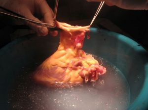
Rejection hurts, but for organ transplant patients it’s more than an emotive issue—it can be a matter of life and death. Having waited months, sometimes years, for a donor and survived major surgery, transplant patients face an uphill battle to prevent their immune systems from rejecting their new organ. Now a new test in which a transplant patient’s blood is scanned for DNA from the donor organ can alert doctors if serious rejection has begun, allowing them to try to stop the process.

Approximately 40% of transplant patients experience at least one episode of acute rejection in the first year after they receive an organ. Catching any major immunological backlash early is key to minimizing its effects, especially because most rejection episodes are reversible with a large dose of immunosuppressant drugs. But patients typically must undergo regular biopsies of their new organ to monitor its health; the procedure is both painful and expensive, and biopsies also risk damaging the organ, explains cardiologist Hannah Valantine of Stanford University School of Medicine in Palo Alto, California. In 2009, Valantine developed a noninvasive rejection test that relies on monitoring a patient’s immune system. AlloMap became the first U.S Food and Drug Administration-approved test for heart transplants, but it still fails to catch about half of rejection events.

To capture the rest, Valantine recently headed back to the drawing board. This time she enlisted help from biophysicist Stephen Quake of Stanford. Together they designed a test that relies on the fact that a transplanted organ’s genome is distinct from that of its new host. The test monitors fragments of DNA released by the organ into the blood, when cells from the transplant tissue are naturally broken down. To validate this strategy, the researchers tried their test on stored blood plasma from organ transplant patients, some of whom had had confirmed rejection episodes. During a rejection event, the levels of circulating DNA from the donor organ go up, making up on average 3% of free DNA in the recipient’s blood rather than the typical 1%, the researchers report online today in the Proceedings of National Academy of Sciences.

Valantine hopes that this test can eliminate the need for regular biopsies as a means of rejection monitoring; patients often have one every month in the first year after a transplant. Instead, physicians would perform confirmation biopsies only if the DNA test results were positive. The new test can detect “very low levels of DNA to predict rejection,” Valantine says, making this approach more sensitive than the AlloMap test. If the test can alert doctors to rejection earlier, she notes, they can “tinker” with the levels of immunosuppressant drugs rather than go in with “a big-gun approach” that lowers the patient’s immune system so much that they are at risk of infection and cancer.

Bruce Rosengard, the surgical director of the cardiac transplantation program at Massachusetts General Hospital in Boston, is cautiously optimistic about the new test. “Organ rejection remains one of the primary obstacles to transplant success,” he says. “Anything that we can do to reduce the number of heart biopsies is a very positive development. … I think this approach will gain traction pretty quickly.”

Valantine hopes to have the new test available to doctors in a year’s time, adding that she sees no reason why it can’t be used to detect rejection of other transplanted organs.

:: Read original here ::

Nosing Out a Smell Gene

The daily smells of the world—the freshness of spring flowers, the smokiness of charred morning toast, the invigorating aroma of a cup of coffee—are lost on those with anosmia, a complete inability to detect odors. Now, thanks to a trio of people who feel no pain, a new study has pinpointed the first set of mutations that are to blame for this condition. The finding could help scientists better understand the gradual loss of smell that happens with age.

The discovery was somewhat happenchance. It began when neurobiologist Frank Zufall of Saarland University in Homburg, Germany, was contacted by pain researcher John Wood of University College London and geneticist Geoffrey Woods of the University of Cambridge in the United Kingdom. Wood and Woods had been studying three people with an inability to feel pain. In 2006, they found that all had mutations in a gene that codes for a sodium channel called Nav1.7 that is involved in the firing of neurons. The researchers also noticed that the volunteers reported having no sense of smell. When the duo created mice with similar mutations, the pups were free of pain, but they also had difficulty smelling; they seemed unable to pick up the odor of their mother’s teats to suckle, for example.

To investigate how the lack of these channels causes anosmia, Zufall and his team studied the genetically engineered mice. They discovered that although neurons in the rodents’ noses respond to different odors, they were unable to transmit these signals to the olfactory bulb, the region at the front of the brain that processes smell.

The researchers also confirmed that the mice couldn’t smell. In a series of experiments, the rodents didn’t avoid the smell of predators, nor did they retrieve their pups when they were scattered around their cage, the researchers report online today in Nature. “This behavior supports that fact that the mice aren’t smelling,” says Lisa Stowers, a neurobiologist at The Scripps Research Institute in San Diego, California, who studies olfaction.

Zufall hopes to turn up more genes underlying anosmia by encouraging doctors to carry out a smell test in patients who present with neurological problems. Peter Mombaerts, an olfaction neuroscientist at the Max Planck Institute of Biophysics in Frankfurt, Germany, agrees that this would be a good approach. “It is a very cheap test,” he says, but he cautions that few doctors currently do it, even though smell can be an important indicator of neurological disease. For example, in Alzheimer’s patients, smell is often one of the first things to deteriorate.

The number of people that are anosmic from birth is likely quite small, says Mombaerts, but by better understanding how olfactory signals are disrupted in people born with this condition, researchers hope to gain insight into why many more develop it later in life.

:: Read original  here ::

How Hormone Puts a Kick in the Sperm’s Tail

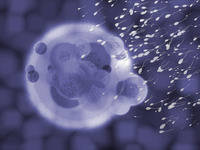
It’s exhausting being a sperm. Having made the long-distance swim up the fallopian tube, a sperm must then rev up its tail to propel itself through the thick jelly-like coating of the egg. The female hormone progesterone, released by the egg, prompts the tail to switch from a smooth swimming motion to a frantic flicking, but exactly how has been puzzling. Researchers have now shown that the hormone acts directly on a sperm surface protein, a discovery that may suggest new nonhormonal contraceptives.

For 10 years, researchers have suspected that progesterone, which the egg releases in huge quantities, is responsible for the asymmetrical, whiplike tail movements that give sperm enough torque to penetrate the ovum. Because sperm respond to progesterone within seconds, scientists reasoned that the hormone must bind to a surface protein and not one within the cells, which would take longer for the progesterone to reach.

In 2001, researchers hoped they had found the progesterone receptor when they discovered that infertile men and mice sometimes had mutations that disrupted a protein, called CatSper, which ferries calcium ions in and out of sperm. This so-called calcium channel is found exclusively within sperms’ tails, but working out whether it responds to progesterone proved a thornier exercise than expected. Sperm are not easy cells to work with—for one thing, they don’t stay still.

Now, two research teams have finally connected progesterone to CatSper by inserting a tiny electrode into individual sperm, a technique usually reserved for measuring the electrical signals in neurons. In independent studies appearing online in Nature today, the groups have documented the change in current inside a sperm as progesterone causes positively charged calcium ions to pass into the cell. And because a working ion channel produces a characteristic electrical fingerprint, the researchers were able to use their electrodes to demonstrate that CatSper was responsible for letting in the calcium.

Such work could ultimately explain why some men whose sperm don’t respond to progesterone have low fertility, says Steve Publicover, a physiologist at the University of Birmingham in the United Kingdom who was not involved in the two studies. Publicover notes that this breakthrough was possible because the teams perfected the electrical monitoring of sperm. Only two or three labs in the world can do this, he confirms.

The findings may prove important for explaining the 40% of male infertility cases for which no underlying cause is known, explains Benjamin Kaupp, a biophysicist at the Center of Advanced European Studies and Research in Bonn, Germany, who led one of the teams. “If we can identify the molecules involved, we can look to see if the cause of a man’s infertility is because one or more of these molecules is not working properly,” he says. Based on this work, for example, clinicians could investigate whether a man’s sperm is insensitive to progesterone due to problems with CatSper.

Polina Lishko, a physiologist at the University of California, San Francisco, who is a member of the other team that made the progesterone-CatSper connection, suggests a different outcome from the research. Current female contraceptives are hormonal, depending on progesterone or estrogen, and cause side effects such as weight gain. Lishko argues that CatSper “offers a great opportunity to develop a nonhormone contraceptive.” Once researchers have located where progesterone binds to the CatSper channel, they can look for molecules that would block this interaction, rendering sperm sterile, she explains. “CatSper channels only occur in the tails of the sperm; [such a] contraceptive would have no effect on females and only disrupt male sperm,” Lishko says.

:: Read original here ::