How bloodsuckers find their blood

Scientists have identified the heat-sensitive facial nerves used by vampire bats to detect their next meal.

The nerves allow bats to pinpoint where the blood flows closest to their prey’s skin so they can feed more efficiently.

Vampire bats are among a handful of animals that use infrared sensors to locate their next meal, but are unique in the way they do it.

The findings are reported in the journal Nature.

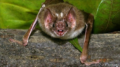
Native to Central and South America, the Common Vampire Bat, Desmodus rotundus, needs to take a sanguineous slurp every night to survive.

Researchers believe that the bats rely solely on detecting their next meal in the dark by listening out for their prey’s breathing.

Having located a prey individual the bats crawls along the ground and onto the animal.

Once atop their prey, the bats are capable of using their heat-adapted nerves in their upper lip and nose to detect blood up to 20cm under their prey’s flesh.

The new finding has pinpointed the molecule that is responsible – heat-sensitive TRPV1. TRPV1, a protein, usually helps animals detect dangerously high temperatures (those over 43 degrees C), but in the bats, some of the TRPV1 molecules have been mutated into a version that is sensitive to lower temperatures, those around 30 degrees C.

Lots of blood-sucking animals search out their next meal using heat-detecting molecules, but they all seem to do it in a different way, said bat biologist, Brock Fenton from the University of Western Ontario, who was not involved in the work.

He said that perceptual world of bats undoubtedly has many more intriguing secrets.

  • The Common Vampire Bat (Desmodus rotundus) is one of three species of vampire bat: The Hairy-legged Vampire Bat (Diphylla ecaudata), and the White-winged Vampire Bat (Diaemus youngi)
  • All three live in the Central and South America
  • D. rotundus feeds mainly on domestic animals, using its razor sharp teeth to make small (5mm) cuts in their prey – most often around the neck or vulva – and secretes an anticoagulant into the wound so it can draw enough blood to the surface
  • D. rotundus drinks its body weight in blood each night, secreting blood plasma in its urine as it feeds to lighten the load
  • Scientists have developed a anti-clotting drug from the saliva of vampire bats that could help stroke patients

:: Read original here ::

Dyslexia makes voices hard to discern, study finds

People with dyslexia struggle to recognise familiar voices, scientists suggest.

The finding is the first tentative evidence that small sounds in the human voice that vary between people are difficult for dyslexics to hear.

Writing in the journal Science, the scientists say that many people could have some degree of “voice blindness”.

And by studying it, scientists hope to better understand how the human brain has evolved to recognise speech.

Humans rely on small sounds called phonemes to tell one person from another.

As we first try to form the word dog, for example, phonemes are the “duh”-“og”-“guh” sounds that our parents prompt us to make.

But as we master the ability to read, we become less reliant on recognising these sounds to read, and eventually stop noticing them.

Despite ignoring them, however, phonemes remain important for voice recognition.

The tiny inflections in the way people pronounce phonemes gives a listener cues to tell one voice from another.

Because people who suffer from dyslexia are known to struggle with phonemes when reading, a US-based team of scientists wondered whether they might also struggle hearing them in people’s voices.

Listen well

To investigate, the team grouped 30 people of similar age, education and IQ into two camps: those with and without a history of dyslexia.

The subjects then went through a training period to learn to associate 10 different voices – half speaking English and half speaking Chinese – with 10 computer-generated avatars.

The subjects were then later quizzed on how many of those voices they could match to the avatars.

Non-dyslexics outperformed people with a history of dyslexia by 40% when listening to English.

However, this advantage disappeared when the groups were listening to Chinese.

Dorothy Bishop from the University of Oxford thinks that this is because “when [they] are listening to Chinese, it is a level playing field, because no one has learned to hear [Chinese] phonemes”.

The researchers think that dyslexics don’t have as comprehensive a phoneme sound library in their heads, and so they struggle when they hear phonemes spoken by unfamiliar voices because their “reference copy” isn’t as well-defined.

“It is a very interesting result. The only thing that I would really like to see to convince me… is if they were to repeat the experiment using Jabberwocky.”

Using Jabberwocky, the nonsense poem by Lewis Carroll, would allow the researchers to determine whether the listeners identify who’s who from the meaning of what they are saying, or whether listeners are purely relying on the phonemes.

Dr Bishop speculated that non-dyslexics may be worse at extracting the meaning of the words, meaning they under perform in this task.

Understanding the mechanics of voice recognition is important, said the study’s lead author Tyler Perrachione from the Massachusetts Institute of Technology in Cambridge, US, because it allows a listener to pinpoint a familiar voice above the hubbub of a crowded room.

Mr Perrachione explained that very little is known about voice blindness, which is formally called phonagnosia.

“In reality, phonagnosia is probably much more common,” he explained, “but people who don’t recognise that voices sound different may not even realise they lack the ability to tell voices apart.”


:: Read original here ::

Age-related brain shrinking is unique to humans

The brains of our closest relatives, unlike our own, do not shrink with age.

The findings suggest that humans are more vulnerable than chimpanzees to age-related diseases because we live relatively longer.

Our longer lifespan is probably an adaptation to having bigger brains, the team suggests in their Proceedings of the National Academy of Sciences paper.

Old age, the results indicate, has evolved to help meet the demands of raising smarter babies.

As we age, our brains get lighter. By 80, the average human brain has lost 15% of its original weight.

People suffering with age-related dementias, such as Alzheimer’s, experience even more shrinkage.

This weight loss is associated with a decline in the delicate finger-like structures of neurons, and in the connections between them.

Alongside this slow decline in its fabric, the brain’s ability to process thoughts and memories and signal to the rest of the body seems to diminish.

Researchers know that certain areas of the brain seem to fare worse; the cerebral cortex, which is involved in higher order thinking, experiences more shrinkage than the cerebellum, which is in charge of motor control.

Yet despite the universality of ageing, scientists do not fully understand why our brains experience this continuous loss of grey matter with age.

Intriguingly, the brains of monkeys do not seem to undergo the same weight loss, raising the question of whether it is a distinctively human condition.

Now, a team of neuroscientists, anthropologists, and primatologists have pooled their expertise and datasets to reveal the answer.

Comparing magnetic resonance images from more than 80 healthy humans between the ages of 22 and 88 with those of a similar number of captive-bred chimps, the researchers found that chimps’ brains do not shrink with age.

The results suggest that the estimated 5-8 million years of evolutionary history that separate chimps from humans have made all the difference in the way that the species age.

It takes a village…

Anthropologist Chet Sherwood from George Washington University in Washington DC, who led the study, thinks that humans live longer to “pay for” their larger-brained children.

Humans live relatively long compared to other great apes. The majority of this extended life is post-menopausal, while chimps are reproductively viable right up to their death.

A human brain is three times the size of chimpanzee’s.

And it is not such a stretch, Dr Sherwood suggests, to conclude that grandparents’ extended lives are in an evolutionary sense there to relieve mothers from being solely responsible for raising their big-brained, energetically costly infants.

“I say this right now, as my seven year old daughter is being looked after by my mother,” he told BBC News.

“Because neurons cannot regenerate, aging, he thinks, is just the stress of living long enough to lend a helping hand to some relatives.”

“[The study] provides very good evidence that the patterns of brain ageing in humans are quite different from other animals,” commented neuroscientist Tom Preuss from Emory University in Atlanta, US, who was not involved in the research.

However, Dr Preuss was clear that these differences do not make other animals useless as models for studying age-related diseases.

Instead, the differences could help to explain why humans suffer more from these diseases than other animals seem to.

:: Read original here ::

Images capture moment brain goes unconscious

For the first time researchers have monitored the brain as it slips into unconsciousness.

The new imaging method detects the waxing and waning of electrical activity in the brain moments after an anaesthetic injection is administered.

As the patient goes under, different parts of the brain seem to be “talking” to each other, a team told the European Anaesthesiology Congress in Amsterdam.

But they caution that more work is needed to understand what is going on.

The technique could ultimately help doctors pinpoint damage in the brains of people suffering from stroke and head injury.

“Our jaws just hit the ground,” said anaesthesiologist Professor Brian Pollard from Manchester Royal Infirmary on seeing the images for the first time.

“I can’t tell you the words we used as it wouldn’t be polite over the phone.”

Although regions of the brain seem to be communicating as “consciousness fades”, Professor Pollard cautions that it is early days and that he and his team from the University of Manchester still have many brain scans to analyse before they can say anything conclusive about what is happening.

The finding supports a theory that is championed by Professor Susan Greenfield, from the University of Oxford, that unconsciousness is a process by which different areas of the brain inhibit each other as the brain shuts down.

The new technique, called Functional Electrical Impedance Tomography by Evoke Response (fEITER), is more compact than other brain imaging techniques, such as functional magnetic resonance imaging (fMRI), and so is easily transported into the operating theatre.

It involves attaching tens of electrodes to the patient’s head, which send low electrical currents through the skull. The currents are interrupted by the brain’s tissues and electrical signals.

Professor Pollard explained that the brain’s structures should not change over a minute-long scan, and so any differences that he and his team see as the patient falls asleep must therefore be due to changes in their brain’s activity.

It is hoped that this technique could be used to learn about the nature of consciousness, but it is also likely to help doctors make headway in monitoring the health of a person’s grey matter after they have suffered a head injury or stroke.

:: Read original here ::

Nosing Out a Smell Gene

The daily smells of the world—the freshness of spring flowers, the smokiness of charred morning toast, the invigorating aroma of a cup of coffee—are lost on those with anosmia, a complete inability to detect odors. Now, thanks to a trio of people who feel no pain, a new study has pinpointed the first set of mutations that are to blame for this condition. The finding could help scientists better understand the gradual loss of smell that happens with age.

The discovery was somewhat happenchance. It began when neurobiologist Frank Zufall of Saarland University in Homburg, Germany, was contacted by pain researcher John Wood of University College London and geneticist Geoffrey Woods of the University of Cambridge in the United Kingdom. Wood and Woods had been studying three people with an inability to feel pain. In 2006, they found that all had mutations in a gene that codes for a sodium channel called Nav1.7 that is involved in the firing of neurons. The researchers also noticed that the volunteers reported having no sense of smell. When the duo created mice with similar mutations, the pups were free of pain, but they also had difficulty smelling; they seemed unable to pick up the odor of their mother’s teats to suckle, for example.

To investigate how the lack of these channels causes anosmia, Zufall and his team studied the genetically engineered mice. They discovered that although neurons in the rodents’ noses respond to different odors, they were unable to transmit these signals to the olfactory bulb, the region at the front of the brain that processes smell.

The researchers also confirmed that the mice couldn’t smell. In a series of experiments, the rodents didn’t avoid the smell of predators, nor did they retrieve their pups when they were scattered around their cage, the researchers report online today in Nature. “This behavior supports that fact that the mice aren’t smelling,” says Lisa Stowers, a neurobiologist at The Scripps Research Institute in San Diego, California, who studies olfaction.

Zufall hopes to turn up more genes underlying anosmia by encouraging doctors to carry out a smell test in patients who present with neurological problems. Peter Mombaerts, an olfaction neuroscientist at the Max Planck Institute of Biophysics in Frankfurt, Germany, agrees that this would be a good approach. “It is a very cheap test,” he says, but he cautions that few doctors currently do it, even though smell can be an important indicator of neurological disease. For example, in Alzheimer’s patients, smell is often one of the first things to deteriorate.

The number of people that are anosmic from birth is likely quite small, says Mombaerts, but by better understanding how olfactory signals are disrupted in people born with this condition, researchers hope to gain insight into why many more develop it later in life.

:: Read original  here ::

Don’t Take That Cookie!

Stop it! Don’t touch that! Sit down and be quiet! Whether you heeded these commands as a child could help predict your future. A new study suggests that people who show less self-control as young children are more likely to have failing health, greater debt, and run-ins with the law later in life.

The idea that willpower is important for success is not new. In the late 1960s, Walter Mischel, a psychologist at Columbia University, tested whether 4-year-old children could resist nibbling Oreo cookies when left alone with a plate of them. He and colleagues found a huge range in willpower, and those children better at resisting the temptation went on to do better in school, scoring higher on the standardized tests. Their parents also judged them to be more attentive, competent, and intelligent. Intrigued, psychologist Terrie Moffitt of Duke University in Durham, North Carolina, and her colleagues sought real-life data to test whether individuals with more willpower and not just self-discipline when offered cookies, achieved greater success in life.

The international team tracked approximately 1000 New Zealand children, born in 1972 or 1973, from the age of 3 years until their early 30s, and another 500 British fraternal twins, born in 1994 or 1995, from the ages of 4 years to 12 years. They used a range of measures to assess the children’s self-control, including their impulsivity, persistence at a task, patience while waiting in line, and hyperactivity.

Compared with their more disciplined twins, children who had less self-control at age 5 were more likely to have begun smoking, performing badly in school, and acting out at age 12, the researchers report online today in the Proceedings of National Academy of Sciences. And these problems continued in later life. Controlling for socioeconomic status and IQ, the researchers determined that people who showed the lowest willpower as children went on to be more than twice as likely to have health problems in their 30s, including high blood pressure, weight problems, lung disease, and sexually transmitted diseases. By the age of 32, they also earned 20% less and were about three times more likely to be dependent on tobacco, alcohol, or harder drugs and to have been convicted of a crime.

Moffitt explains that people didn’t fall into two categories—disciplined or undisciplined—but existed on a spectrum. “It means all of us could benefit from improving our self-control,” she says.

Moshe Bar, a psychologist at Harvard Medical School in Boston who was not involved in the study, is impressed by the long-term data set but cautions that the study is observational and can’t establish that self-control breeds success. Still, parents of cookie nibblers shouldn’t despair, he says. He was intrigued to read that some of the children in the study improved their self-control. Bar and his 7- and 10-year-old children “play a game of ‘waiting’ with unwrapping a candy for no other reason than practicing a delay,” he says. “It works,” he says, but then, as he points out, he has a sample size of only two.

:: Read full here ::

U.K. Neuroscientists Complain Funding Cut Penalizes Them for Success

LONDON—At a briefing here today, the British Neuroscience Association (BNA) warned that a planned 20% cut in funding for neuroscience by the Biotechnology and Biological Sciences Research Council (BBSRC) will drive an estimated 100 researchers in the field from the United Kingdom and weaken the nation in a scientific discipline in which it has traditionally excelled. In a letter addressed to Tom Blundell, chair of the council of the BBSRC, which more than 100 neuroscientists put their name to, BNA protested the funding cut and called for its reconsideration.

In January, BBSRC revealed its plans to prioritize particular research disciplines that it believes will help address major challenges to future society. BBSRC’s favored themes include: food security, bioenergy and industrial biotechnology and basic bioscience underpinning health and wellbeing. The Council said that meant that neuroscience, which currently accounts for 13% of grant funding (amounting to £150 million), will receive less funding. BBSRC’s head Douglas Kell recently defended the cut in a blog post.

BNA says that BBSRC’s reprioritization smacks of a desperate measure to balance the books, and that the field of neuroscience is being penalized for being successful. British neuroscientists contend that they have a great track record, helping to explain brain illnesses such as motor neuron, Parkinson’s, and Alzheimer’s diseases, and develop many important drugs such as Fluoxetine, the widely used antidepressant known as Prozac. Eli Lilly, a U.S.-based pharmaceutical company, found that neuroscience research in the south of England was the most cited globally.

David Nutt, a neuropsychopharmacologist at Imperial College London says that there has been no explanation by BBSRC for their dramatic change in strategy, and no consultation on these cuts. The effects will be felt immediately, he says. It just seems like an “arbitrary exclusion.” “I don’t know what to tell my PhD students … I don’t see jobs for them,” he remarks.

Duncan Banks, the director of BNA , argued that the BBSRC’s funding cut, amounting to about £4 million in spending on neuroscience, will particularly affect opportunities for young scientists, he explains.

BBSRC cuts come at a time when the funding for basic neuroscience in the United Kingdom is already threatened with a move by the Medical Research Council to fund more clinically relevant science, explains Colin Blakemore, a neuroscientist at the University of Oxford and former head of the Medical Research Council. Yet one-third of global health issues are related to neurological problems, such as depression, pain, and the diseases of the aging brain. BNA points out that what really drives new treatments is basic research. Blakemore remarks, “Neuroscience remains one of the least understood areas of biology.” “An aging population should be very worried about cuts in neuroscience,” he says.

The strength of the U.K.’s basic research was what originally attracted pharmaceutical companies to the United Kingdom, notes Blakemore. Yet that attraction may be fading, especially in the neuroscience. Recent closures of many pharma-funded institutes working in the field of neuroscience—Pfizer, GSK, Astra Zenica, and Merck have all closed institutes in the last year—may also force neuroscientists out of the United Kingdom.

:: Read original here ::

A Woman’s Tears: The Anti-Viagra?

For many a man, few things deflate his passion faster than the sight of a woman crying. But tears may do more than visually tell a man it’s not time for romance. A woman’s tears contain substances that reduce men’s sexual arousal, a new study indicates. It’s the first evidence that human tears contain chemical signals.

Tears have largely been considered just a visual signal among people: Studies have shown that people looking at a sad face perceive it as sadder when tears are added. In contrast, some animals seem to use their waterworks to communicate chemically. The tears of male mice, for example, contain a protein that makes females more receptive to mating. But given that people, unlike rodents, don’t preen each other, researchers assumed that we rarely come in close enough contact to perceive chemical cues in tears.

Noam Sobel, a neurobiologist at the Weizmann Institute of Science in Rehovot, Israel, wasn’t convinced. Human tears shed under duress differ chemically from those shed to clear the eye of irritants, and he wondered whether human tears might also carry messages for the opposite sex.

To find out, he recruited two women who claimed they could cry on demand. He showed them a sad film—a scene from Franco Zeffirelli’s 1979 film The Champ, in which a son cries over the body of his dying father, a boxer—and collected their tears in vials (see video). Within minutes, the vials were handed to 24 men, aged 23 to 32, who took 10 deep breaths over the open receptacles. Researchers also stuck a tear-soaked cotton square under each man’s nose for the duration of the experiment. As a control, Sobel and his team did the same with saline solution, which they trickled down the women’s cheeks to account for perfumes and face creams they might have been wearing.

The men were then asked to judge the emotion and attractiveness of images of women’s faces that had been made emotionally ambiguous by morphing together happy and sad faces. The men couldn’t smell the difference between tears and saline, and the tears did not influence how sad they thought the faces seemed. Nevertheless, the men found the women less attractive after smelling the tears, the researchers report online today in Science.

The men’s heart and breathing rates, skin temperature, and testosterone levels also sank, indicating a drop in sexual arousal. Peering into the subjects’ brains using functional magnetic resonance imaging, the researchers found that on average the regions of the brain that usually light up when an individual is aroused, the hypothalamus and fusiform gyrus, responded normally to moderately erotic images. However, this neural activity was dampened when the men were exposed to the tears.

Shedding tears is just another way, along with pheromones and body language, that the sexes can communicate, says Sobel. Women shed tears significantly more often during menstruation, when there is a low chance of conceiving, he notes. “This makes perfect sense because it is signaling that sexual activity is inappropriate from an evolutionary point of view,” says Sobel.

The study’s results expose “a hidden, underlying origin” for tears, says Adam Anderson, a neuroscientist at the University of Toronto in Canada. Tears originally functioned to simply shed irritants from the eyes. They were then co-opted to contain chemosignals and then perhaps further co-opted to express sadness, Anderson says.

But many questions remain, says Kazushige Touhara, a molecular biologist at the University of Tokyo who works on biochemical signaling among mice. The substances that dampen male arousal remain unidentified, and it’s not clear whether the men sensed them with their olfactory system or through their skin. Without knowing such details, says Touhara, it is difficult to know how important tears are as a biochemical signaling route in human interactions.

:: Read original here ::

Do Chimps Play With Dolls?

In the tropical rainforest of Uganda’s Kibale National Park, a young female chimpanzee seems to have adopted a stick. She’s holding it close to her abdomen and carrying it with her everywhere she goes. In a new study, the first to document this behavior in the wild, researchers argue that stick cradling may be akin to human children playing with dolls. And because the team observed it far more frequently in female chimps, the findings suggest that certain gender-specific behaviors are hard-wired.

Making the observations was no easy task. Harvard University primatologist Richard Wrangham and colleagues spent 12 hours a day—much of it crouched behind vegetation—tracking a group of 68 chimps through thick rainforest. During the study period, they observed about 300 instances of chimps picking up sticks. In 40% of cases, the chimps cradled the sticks in the crook of their arm or tucked between their abdomen and thigh (see picture), whereas the rest of the time they used sticks to probe trees and fight each other. More than three-quarters of the stick cradlers were female, the team reports online today in Current Biology. Females were also 10 times more likely to use the sticks as tools than males were—the first time such a large disparity has been reported.

Wrangham says stick cradling reminded him of doll play in girls. And because chimp mothers don’t play with sticks, young female chimps probably aren’t learning the behavior from watching their moms. Instead, it may be hard-wired. Given the close evolutionary relationship between chimps and humans, the implication is that doll playing and other gender-specific behaviors seen in young human children may be hard-wired as well, he says.

Rebecca Jordan-Young isn’t willing to go that far. A sociomedical scientist who studies sex, gender, and sexuality at Barnard College in New York City, Jordan-Young says she doesn’t think anyone should draw sweeping interpretations about innate differences between the sexes from this study. She says that the researchers cannot rule out that females carry sticks because they are mimicking the behavior of other adolescent females. It could also just be a cultural fad.

In addition, Jordan-Young questions the emphasis the authors placed on the doll-carrying observations. Just as intriguing, she says, is the fact that female chimps use tools much more often than males do. But those types of findings, she says, don’t grab headlines.

:: Read original here ::