Monkeys Chew the Cud

Eating your greens can be grueling, especially if you are a monkey who dines on a high fiber diet. Some primates overcome their digestive dilemma by hosting microbes in their guts that help them breakdown the tougher leaves, much like cows do. Cows and other ruminants also maximize this symbiotic relationship by regurgitating and rechewing their stomach contents to get the most out of each meal. This behavior was considered unique to four-legged herbivores. Now researchers have witnessed proboscis monkeys that live in the mangroves and swamps of Borneo doing the same (see the video). This is the first evidence that primates ruminate, too, reports the team online today in Biology Letters, and gives us all something to chew over.

:: Read original here ::

The Oldest Buttercup Yet

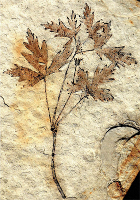
Darwin called the origin of flowering plants an “abominable mystery.” They appear in the fossil record and immediately grow abundant and varied, creating a problem for his theory of slow but continuous change. The unearthing of a new fossil in northeast China, described online today in Nature, could explain the apparent contradiction. The ancient flowering plant, Archaefructus liaoningensis, resembles a modern-day buttercup, with slender stems and three-lobed leaves. Its discovery pushes back the date of when flowering plants diversified to around 127 million years ago, during the early Cretaceous period. That’s a couple of million years earlier than Darwin had previously thought, suggesting that these ancient blooms had longer to evolve than he suspected.

:: Read original here ::

Worms Are Divided After All

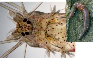
It turns out that worms really are deeply divided. In the mid-19th century, French naturalist Jean Louis Armand de Quatrefages de Bréau split worms into wigglers—which crawl and swim to their hearts’ content, such as the marine ragworm (left)—and the more sedentary, typically tube-dwelling nonwigglers, such as the earthworm (right). However, early genetic studies called this classification into question. They indicated, for example, that the wigglers weren’t that closely related to one another; instead, their physical similarities were the result of their adapting to similar lifestyles and environments. Now more comprehensive genetic evidence, reported online today in Nature, shows that de Bréau was right after all. Analyzing 231 genes from 34 different annelids, otherwise knows as ringed worms, researchers have shown that wigglers and nonwigglers do indeed represent two different evolutionary groups. What’s more, the team found that the split between the two groups happened very early in worm evolution; the exact date isn’t known because the soft bodies of worms don’t preserve well as fossils. Wigglers kept their bristled appendages for moving around and foraging, whereas nonwigglers lost them as they evolved to stay put in their burrows to eat sediments and plankton.

:: Read original here ::

Whistling Caterpillars Shake Off Predators

When in danger, whistle. It works for the walnut sphinx caterpillar (Amorpha juglandis). The fat, juicy larvae of butterflies and moths tend to be experts at predator avoidance, using camouflage, rolling themselves in leaves, and even flicking their own poop to discourage birds, frogs, and small mammals from eating them. Whistling is just another string in their bow, researchers report online this week in The Journal of Experimental Biology. When the team used forceps to simulate the peck of a bird’s beak, the caterpillars forced air through the small holes on either side of their body—normally used for breathing—to produce a high-pitched whistle. When yellow warblers heard the noise, these natural enemies of the caterpillars hesitated, jumped back, or flew off. The sound may have startled them, or perhaps they found the tune indigestible.

:: Read original here ::

Rethinking Brain Evolution in Insects

As surprising at it may seem, wasps, bees, and even ants have relatively large and complex brains. That allows these “social insects” to keep track of the intricate relationships between the thousands of individuals in their colony—or so researchers thought. A new study indicates that these insects didn’t grow big brains to cope with social living; they evolved them millions of years earlier when they were solitary parasites.

The link between brain size and social living was first noted in 1850, when scientists identified mushroom bodies in the insect brain. Aptly named because they’re shaped like mushrooms, the structures contain thousands of neurons responsible for processing and remembering smells and sights. Social insects tend to have larger mushroom bodies than solitary ones, leading researchers to believe that the transition from solitary to social living increased the size of these brain regions.

But Sarah Farris has found a different explanation. Instead of comparing social insects with solitary ones, Farris, a neurobiologist at West Virginia University in Morgantown, looked into the past. To get a sense of how the wasp brain evolved over time, she and taxonomist Susanne Schulmeister of the American Museum of Natural History in New York City compared the mushroom bodies of parasitic wasps with those of nonparasitic wasps, which represent the very oldest form of wasp. The parasitic wasps had consistently larger and more elaborate mushroom bodies than the nonparasites, the duo reports online today in the Proceedings of the Royal Society B. In particular, the caps, called calyces, of the parasitic mushroom bodies were twice the size of nonparasites.

Farris points out that parasitism evolved 90 million years before social insects appear, and so “insects had big mushroom bodies for quite a while before sociality arose.” This is the first evidence that parasitism, and not sociality, was the driver of insect mushroom body complexity, she says. That may be because well-developed mushroom bodies help parasitic wasps better locate the nests of the larvae they lay their eggs in.

Francis Ratnieks, an evolutionary biologist at the University of Sussex in the United Kingdom agrees with the study’s findings, but he thinks the researchers need to also look at the brains of social insects. It would be useful, for example, to compare the brains of social worker bees, which process vast quantities of visual information as they fly from flower to flower, with those of parasitic wasps. If bees have even larger mushroom bodies than parasitic wasps, he says, this would suggest that social insects have further improved on the brains that they inherited from their ancestors.

:: Read original here ::

Ancient Tweaking


Twenty years ago, scientists knew nothing of the scraps of RNA that are now known to influence just about every process in our bodies. Back then, the textbooks were simpler: genes code for proteins via the intermediate of RNA, and proteins called transcription factors regulate other proteins. This recipe was so entrenched in the basic orthodoxy of molecular biology that it was even given the name ‘the central dogma’ by the co-discoverer of DNA, Francis Crick.

Scientists now know, however, that this classic view of protein regulation is far too blunderingly inefficient for evolution to settle for. At some point hundreds of millions of years ago, the generation of a small stretch of RNA that could tweak this process gave an individual the edge over everyone else. And so regulatory RNA was born. These scraps of RNA – on average only 22 nucleotides long and now dubbed microRNAs, or miRNAs for short – bind to some messenger RNAs and label them for inactivation or destruction.

So far thousands of miRNAs have been identified in animals. These superintendents of protein regulation are involved in the earliest stages of an animal’s development, determining which cell types grow where and when, and how these cells differentiate into the different body parts. However, since the discovery of miRNAs, many scientists have wondered whether the same miRNAs govern specific tissues in different animals. Knowing this would not only give clues to the age of these different miRNAs, but
also to the age of the cells in which they are found.

:: Read more here ::

Arsenic-munching bacteria found

Arsenic-munching bacteria

In the warm, bubbling pools of Mono Lake in California, scientists have isolated a bacterium that fuels itself on arsenic.

Combining light and arsenic, these bacteria make their food and multiply using a chemical that is toxic to most other life forms.

The researchers think using arsenic as an energy source was a process used by ancient bacteria.

Their findings are reported in the journal Science.

Ronald Oremland of the US Geological Survey explained that these bacteria are photosynthetic, using sunlight – like plants – to turn carbon dioxide into food.

What is different about them is that instead of using water in this process, they use arsenic.

The US-based researchers isolated the bacterium from the lake, which lies at the foot of the Sierra Nevada.

Colour film

“These lakes are fed by hydrothermal waters that leach out arsenic-containing minerals from the surrounding rocks,” Dr Oremland told BBC News.

The researchers noticed that the bacteria had colonised small, hot pools, forming colourful “biofilms”.

MonoLake(USGS)

Bacteria living in Mono Lake, California can survive the high levels of arsenic

“We suspected that these bacteria were using arsenic to make a living, so we scraped the biofilms off the rock and studied them under laboratory conditions.”

By first withholding light, then arsenic, the team showed that the bacteria required both to grow.

This the first time an organism has been found that can use arsenic to photosynthesise under anaerobic conditions, Dr Oremland believes.

He suspects that this is an ancient ability in bacteria.

“We think that bacteria were photosynthesising before oxygen was present in the atmosphere,” he said.

Primordial niche

Understanding how arsenic is metabolised by bacteria could help scientists comprehend its damaging effects inside human cells.

Worldwide, 144 million people are exposed to toxic levels of arsenic in their drinking water.

It enters the body’s cells by diffusion; and once inside, it disrupts how they function by binding to their machinery, inactivating it, and disrupting the way energy is transported.

Long-term exposure can lead to skin disease and kidney and bladder cancer, and it is thought to stunt the intellectual development of children.

The most arsenic-contaminated regions are in India, Pakistan, and China, where soluble arsenic in ground waters is above the World Health Organization’s (WHO) suggested maximum safe level of 10 parts per billion.

:: Read original here ::