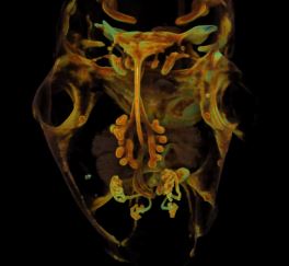
 If you ever wanted to know how the inner organs of a mouse embryo form, this is the movie for you. The animation was created by imaging thin sections of an embryo and then stacking these images to make a 3D movie. “If you look closely, you can see the developing lungs, gut, kidney, and bladder,” says the movie’s creator, Ian Smyth, a developmental biologist at Monash University in Australia. His video was selected from among several for being the most “striking and technically excellent” of the animations submitted to this year’s Wellcome Image Awards in London. Smyth uses these animations to compare normal tissues in embryos with those whose development is disrupted because of disease or exposure to a toxin. The animation was selected because of its ability to illustrate how effective this imaging technique can be for looking at the internal structure of the organs in a noninvasive way, explains Catherine Draycott, one of this year’s judges. “You can almost travel through [the mouse] as it develops.”
:: Read original here ::