Why Most Snails Coil to the Right

sn-snails
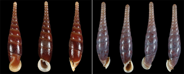
GUELPH, CANADA—When plucking a snail from the beach you’d be lucky to snag a left-coiling shell. That’s because only 5% of all snails are “lefties,” new research shows. Shell enthusiasts have long marveled at the lack of sinistral (left-coiling) snails among their collections, especially when other shelled mollusks, such as clams and the now-extinct ammonites—nautiluslike creatures that sported dozens of tentacles inside spiraled shells—are just as likely to be left- as right-coiling. Now, in the largest survey of its kind, researchers inspected more than 55,000 snail species—representing two-thirds of all gastropods—to reveal that left-coiling has arisen more than 100 times, and yet few of the species that have made the switch have been particularly successful. In the rare cases where left-coiling took off, it was almost always on land, the team reported here in a presentation last week at the annual meeting of the Canadian Society of Zoologists. The researchers don’t know why sinistrality is so rare underwater, but the most likely explanation, they say, is that unlike land snails that tend to hang around where they hatch out, the microscopic young of sea snails are carried on ocean currents that make the chance of meeting and reproducing with another left-coiling nest-mate slim. Without such a meeting, the left-coiling lineage goes extinct.

:: Read more here ::

Multicellularity Driven by Bacteria

F1.smallMONTREAL, CANADA—When taking a dip this summer you will probably swallow tens, possibly hundreds, of microscopic plankton called choanoflagellates. These common organisms have led to an uncommon insight into how multicellular organisms might have evolved. Bacteria can prompt single-celled choanoflagellates to divide into multicellular versions of themselves, University of California (UC), Berkeley, biologist Nicole King reported last week here at the 71st annual meeting of the Society for Developmental Biology. King hopes the work will prompt biologists to look more closely at the role of microorganisms in the evolution of multicellularity.

To the untrained eye, choanoflagellates look like animals. But they are less complex—the closest living relatives of animals but on an older branch of the tree of life. As such, these organisms can provide clues about what early animals looked like and can help reconstruct the events from more than 600 million years ago that led to the incredible diversity of the animal kingdom.

To investigate the transition to colony life, King decided to sequence the genome of a colony-forming choanoflagellate and compare it with the genome of a unicellular individual. But before sequencing, she asked undergraduate Richard Zuzow to purge the sample of everything but the plankton itself. When Zuzow added antibiotics to get rid of any bacteria, the choanoflagellate colonies disappeared. At first, “I didn’t believe him,” King recalls. But with repeated tests, she became convinced that “the bacteria are the important part of the [multicellular] story,” she says

:: Read more here (behind pay-wall) or download pdf here ::

A rare he-she butterfly is born in London’s NHM

A half-male, half-female butterfly has hatched at London’s Natural History Museum.

A line down the insect’s middle marks the division between its male side and its more colourful female side.

Failure of the butterfly’s sex chromosomes to separate during fertilisation is behind this rare sexual chimera.

Once it has lived out its month-long life, the butterfly will join the museum’s collection.

Only 0.01% of hatching butterflies are gynandromorphs; the technical term for these strange asymmetrical creatures.

“So you can understand why I was bouncing off of the walls when I learned that… [it] had emerged in the puparium,” said butterfly enthusiast Luke Brown from London’s Natural History Museum.

Mr Brown built his first butterfly house when he was seven, and has hatched out over 300 thousand butterflies; this is only his third gynandromorph.

Half and half

It is not only the wings that are affected, he explained. The butterfly’s body is split in two, its sexual organs are half and half, and even its antennae are different lengths.

“It is a complete split; part-male, part-female… welded together inside,” he told the BBC.

The dual-sex butterfly is an example of a Great Mormon, Papilio memnon –a species that is native to Asia.

With a shortage of butterfly-specific gender neutral pronouns, the butterfly is being referred to as “it”, and is already middle-aged at three and a half week’s old.

So the public has only a narrow window of opportunity to see it alive.

Though rare, gynandromorphy isn’t unique to butterflies; individual crabs, lobsters, spiders and chickens have all been found with a mix of two sexes.

There are likely many more cases in the natural world, but sexual chimeras are more difficult to spot in animals where females and males look alike.

:: Read original here ::

Researchers Grow Protoeye in Dish

It’s not quite Avatar, but a movie released today shows, in three dimensions, how a chaotic patch of cells grows into the beginnings of an eyeball. Even more impressive, this protoeye was grown in a laboratory dish. Scientists say the finding brings them closer to growing entire organs in the lab, including eyes that could replace those damaged by injury or disease.

In the past decade, researchers have made dramatic progress in understanding how eyes form. They have learned how to turn on and off essential genes and how to transform embryonic stem cells into retinal cells, which can be transplanted into mice to restore vision. But so far, growing an entire eyeball in the lab has eluded scientists, largely because they’ve been unable to recreate the “optic cup,” a chalice-shaped structure that becomes the back of the eye.

Now Yoshiki Sasai of the RIKEN Center for Developmental Biology in Kobe, Japan, and his colleagues have induced embryonic mouse stem cells to spontaneously form the optic cup in a dish. The key ingredient was a mixture of jellylike proteins, called Matrigel, which forms an enticing bed on which stem cells seem to prefer to lie before turning into the eye’s various structures.

In the movie, the cells—made to glow green—push out before inverting and forming two different layers: the first, the retinal cells, and the second, the neurons. This is the first demonstration that stem cells direct their own development in the eye, the team reports online today in Nature.

Knowing that stem cells can direct their own development is key if we want to grow organs without having to also grow the tissues that usually develop around them, says developmental biologist Jane Sowden of University College London, who was not involved in the study. She says that even though Sasai’s team hasn’t yet grown an entire eyeball in a dish, the work shows that it’s possible to grow specific eye structures, such as retinas, from stem cells in a great enough quantity that they could be used in therapy. If the researchers can get the technique to work with human stem cells, she says, it could help the one in 3000 people born with a form of blindness caused by damaged retinal cells and the many more who lose their sight because of age-related disease.

:: Read original  here ::

The Secret of the Glowing Snail


Talk about an inner glow. The marine snail, Hinea brasiliana, radiates green light to startle predators, so the snail can make a quick—or at least relatively quick—get away. But there’s a mystery to this bioluminescence: The snail’s body sports just a handful of glowing cells, yet its entire shell lights up. To shed light on the puzzle, researchers shed some light on the snails. They focused a tight beam of light through the shell’s opening, mimicking the light emitted from the animal’s cells, and found that the entire snail lit up. The trick appears to be that the mollusk’s shell scatters light. This allows the snail to turn a tiny glow into a much larger one, making it seem more formidable to predators. Understanding how the shell’s internal structure produces this luminosity could inspire lighting designs of the future, the team reports online today in the Proceedings of the Royal Society B.

:: Read original here ::

The Curious Case of the Backwardly Aging Mouse

In F. Scott Fitzgerald’s short story, “The Curious Case of Benjamin Button,” an old man gets younger with each passing day, a fantastic concept recently brought to life on film by Brad Pitt. In a lab in Boston, a research team has used genetic engineering to accomplish something similarly curious, turning frail-looking mice into younger versions of themselves by stimulating the regeneration of certain tissues. The study helps explain why certain organs and tissues break down with age and, researchers say, offers hope that one day such age-related deterioration can be thwarted and even reversed.

As we age, many of our cells stop dividing. Our organs, no longer able to rejuvenate themselves, slowly fail. Scientists don’t fully understand what triggers this, but many researchers suspect the gradual shrinking of telomeres, the protective DNA caps on the end of chromosomes. A little bit of telomere is lost each time a cell divides, and telomerase, the enzyme that maintains caps, isn’t typically active in adult tissues. Another piece of evidence: People with longer telomeres tend to live longer, healthier lives, whereas those with shorter telomeres suffer more from age-related diseases, such as diabetes, Alzheimer’s, and heart disease.

Several years ago, Ronald DePinho, molecular biologist and director of the Belfer Institute of the Dana-Farber Cancer Institute, and colleagues at Harvard Medical School in Boston genetically engineered mice to lack a working copy of the telomerase gene. The animals died at about 6 months—that’s young for mice, which usually live until they are about 3 years old—and seemed to age prematurely. At an early age, their livers and spleens withered, their brains shrank, and they became infertile. By early adulthood, these mice exhibited many of the maladies seen in 80-year-old humans.

DePinho says he wondered what would happen to the aging process in these mice if they suddenly began making telomerase again. “Would [we] slow it, stabilize it, or would we reverse it?” He and his colleagues genetically engineered a new batch of mice with the same infirmity, but this time they added back a telomerase gene that became active only when the mice received a certain drug. The researchers kept the gene off during development and let these mice prematurely age, as the previous ones had. But then at 6 months, the team switched on the telomerase gene.

The burst of telomerase production spurred almost total recovery. The rodents became fertile, their livers and spleens increased in size, and new neurons appeared in their brains, the researchers reported online yesterday in Nature.

The ability to reverse age deterioration in the mutant mice indicates that the cells that divide to replenish tissues don’t simply die when their telomere clock expires, says DePinho. They apparently persist in a dormant state from which they can be revived. “One could imagine applying this approach to humans,” he says, focusing the therapy on specific tissue types such as the liver, where telomerase is thought to play an important role in regeneration after damage by hepatitis, parasitic infection, and alcoholism.

K. Lenhard Rudolph, who studies stem cell aging at the University of Ulm in Germany, says that the results are encouraging for people with diseases that cause accelerated aging, like progeria, because the mice in this study were rescued despite already suffering from the effects of chronic disease. “It is a proof of principle that telomeres are at work here.”

Drug companies and researchers are seeking ways to restore, protect, or extend a person’s telomeres, but the jury is still out on whether such interventions can slow the symptoms of aging, let alone reverse them. Telomere investigator Maria Blasco of the Spanish National Cancer Research Center in Madrid cautions that DePinho’s experiment shouldn’t raise people’s expectations of antiaging therapies just yet. “This study uses genetically modified mice,” she says. “What remains a very important question in the field is can you delay aging in a normal mouse?”

DePinho agrees with those concerns. He also warns that his approach has potential drawbacks, as increasing telomerase activity beyond its natural levels can cause cancer. Still, that may not be an insurmountable problem if telomerase levels can be carefully controlled. DePinho notes that the mice in his study, whose telomerase activity was returned to a natural level, didn’t develop tumors.

:: Read original here ::

Ancient Tweaking


Twenty years ago, scientists knew nothing of the scraps of RNA that are now known to influence just about every process in our bodies. Back then, the textbooks were simpler: genes code for proteins via the intermediate of RNA, and proteins called transcription factors regulate other proteins. This recipe was so entrenched in the basic orthodoxy of molecular biology that it was even given the name ‘the central dogma’ by the co-discoverer of DNA, Francis Crick.

Scientists now know, however, that this classic view of protein regulation is far too blunderingly inefficient for evolution to settle for. At some point hundreds of millions of years ago, the generation of a small stretch of RNA that could tweak this process gave an individual the edge over everyone else. And so regulatory RNA was born. These scraps of RNA – on average only 22 nucleotides long and now dubbed microRNAs, or miRNAs for short – bind to some messenger RNAs and label them for inactivation or destruction.

So far thousands of miRNAs have been identified in animals. These superintendents of protein regulation are involved in the earliest stages of an animal’s development, determining which cell types grow where and when, and how these cells differentiate into the different body parts. However, since the discovery of miRNAs, many scientists have wondered whether the same miRNAs govern specific tissues in different animals. Knowing this would not only give clues to the age of these different miRNAs, but
also to the age of the cells in which they are found.

:: Read more here ::

Aberrant appendages

Having a second pair of hands might seem like an advantage but animals born with extra limbs, because of changes in their DNA, generally do not fair well. For more than 25 years, scientists have known about the existence of a mutation in a fruit fly gene that causes just such aberrant appendages, yet the identity of this
gene remained a mystery.

That is until developmental biologist Jürg Müller and his team at EMBL Heidelberg set out to find the gene responsible. By comparing the DNA of mutant and normal flies, Jürg’s group pinpointed the mutation and found that it disrupts the genetic code for the protein Ogt, an enzyme that sticks sugar molecules to the outside of proteins.

:: Read more here ::

Pinning the tail on the histone

Nearly 60 years ago, Pamela Lewis, a geneticist at the California Institute of Technology in Pasadena, noticed that some of the flies she was experimenting on had tiny comb-like structures on their second and third pairs of legs, and not
just the first pair as is usual.

Lewis called these structures ‘sex-combs‘ because males use them to grasp females during mating and she went on to discover the first Polycomb gene, one of many such genes now known to encode proteins that disrupt head to-tail body patterning in a variety of animals, ranging from humans to fruit flies to worms.

:: Read more here ::