Cull ‘cannot save’ Tasmanian devil

Culling does not effectively control the contagious cancer threatening the Tasmanian devil, a new study suggests.

The researchers modelled the effect of removing sick animals on the disease’s prevalence in a small population.

The study, in the Journal of Applied Ecology, seems to confirm findings in wild trials, that selective culling of sick animals is ineffectual at stopping the spread of the disease.

All trial culls of the devils have now been stopped.

Culling has been used to control infectious diseases in a range of species from deer to badgers, wolves to domestic cattle.

Despite proving successful in controlling the diseases of livestock, such as foot and mouth, culling wild animals is controversial because of the lack of evidence that it works.

In fact, cases exists where culling wild animals has made the problem worse.

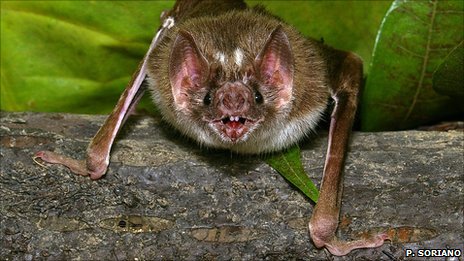
But, hoping to save the Tasmanian devil, Sarcophilus harrisii, from the facial cancer that has wiped out more than 90% of individuals in some areas, conservation biologists have trialled a cull since 2004.

Costly cull

As part of the trial cull, researchers have trapped and euthanised sick animals two to five times a year from an isolated population in the south-east of Tasmania.

Each year, the project costs more than $200,000 (£122,000). Critics say this money could otherwise be spent on captive breeding programmes.

To assess the impact of the cull, Australian researchers Nick Beeton from University of Tasmania and Hamish McCallum from Griffith University created a computer model in which they simulated the effects of the cull.

“We found the removal rate required to suppress disease was higher than that which would be feasible in the field,” explained Mr Beeton.

In the field, biologists find that 20% of the population is never captured and could be acting as a reservoir for the disease.

Unless all the devils are trapped and inspected, including the “trap-shy” ones, continuous culling is unlikely to be an effective disease control, the researchers write.

Mr Beeton told BBC News: “The disease suppression trial was ended as this paper was being written.”

“Our research demonstrates that we must be flexible and be prepared to change strategy as new information comes to light,” he added.

“It’s much better to do a study like this, than spend a lot of money on a huge culling programme and then find that it hasn’t worked,” said geneticist Elizabeth Murchison from the Welcome Trust Sanger Institute in Hinxton, UK, who studies the devil cancer.

She added that by confirming that culling does not work, conservationists can then focus their efforts on alternative strategies, such as the captive breeding programme and developing a vaccine against the cancer.

The transmissible facial cancer

  • Devil Facial Tumour Disease (DFTD) is spread by biting during aggressive encounters
  • The living cancer exists as contagious clone cells; highly unusual for a cancer. There is only one other transmissible cancer known, which affects dog genitals
  • The devil’s immune system seems unable to detect the cancer
  • The disease forms tumours around the mouth interfering with feeding, leading to death
  • The cancer originally arose in Schwann cells – cells which wrap themselves around nerve tissue
  • First seen in 1996, the cancer has badly hit Tasmanian devil populations

:: Read original here ::

Is It Folly to Take Folic Acid?

For mothers-to-be, doctors worldwide advise taking a folic acid supplement. That’s because pregnant women with a deficiency of this vitamin have an increased chance of giving birth to a baby with serious birth defects, such as spina bifida and anencephaly. Yet a new mouse study shows that folic acid supplementation can itself sometimes increase the risk of birth defects or even cause the death of embryos. Experts caution, however, that the unexpected rodent results are too preliminary to require an immediate change in medical practices until more is known about how the vitamin influences development.

People typically obtain folic acid, or folate, from consuming leafy vegetables, but not everyone gets enough from their diet, particularly pregnant women. The vitamin plays a key role in the development of the neural tube, the embryonic region that gives rise to the spinal cord and brain.

Evidence from randomized clinical trials has shown that babies born to women who double their recommended daily dose of folic acid are between 40% and 50% less likely to have birth defects of the spine, skull, and brain. As a result, the United States has fortified most of its grains with this vitamin since 1998, and a handful of other countries have followed suit.

To the surprise of the researchers, in three of the five strains, the extra folic acid seemed to worsen the severity of birth defects rather than remedy them. In one of the mutant lines, dubbed L3P, eating the higher folic acid diet long term increased the chance that young were born with neural tube defects from 20% to 60%, the group reports in the 15 September issue of Human Molecular Genetics. And for another strain, Shroom3, many of whose embryos don’t naturally survive until birth due to their genetic problems, eating the higher folic acid food significantly increased the percentage of lost embryos.

Niswander says it is clear that folic acid is good for human fetuses, but the new study makes her wonder whether high levels of the vitamin may be harmful in some circumstances. Still, she stresses that more data are needed before any serious reconsideration of how much folic acid to recommend for impending mothers.

Roy Pitkin, a retired University of California, Los Angeles, researcher who specialized in pregnant women’s nutrition and chaired an Institute of Medicine panel that in 2000 reviewed folic acid’s health effects also cautions against a rush to judgment: “It would really be throwing the baby out with the bath water to say that because of this one mouse study, we are going to question the food fortification.”

Especially, he says, because we know that species differ considerably from each other when it comes to birth defects. He offers the example of thalidomide—a drug given to pregnant women in the 1950s to cure morning sickness—that causes severe human birth defects but that is perfectly safe in rats.

Neuroscientist Elizabeth Grove of the University of Chicago studies how the mammalian brain develops and echoes this concern. She warns that researchers have found hundreds of mutations that cause birth defects in mice but that so far don’t seem to produce the same problems in humans. The effects of folic acid may also be species specific, she says.

:: Read original here ::

Swedish wolves threatened by under-reported poaching

Illegal poaching accounts for over half of all deaths of Swedish wolves, suggests a new study.

Basing their estimates on long-term wolf counts, the researchers reveal that two-thirds of poaching goes undetected.

The study suggests that without the past decade of persecution Swedish wolves would be four times more abundant than they are today.

The study’s findings are reported in Proceedings of the Royal Society B.

“Many have speculated that poaching levels are high for many threatened species of carnivores,” said Chris Carbone from the Zoological Society of London.

“This study presents an important step in trying to quantify this hidden threat,” he added.

The new study predicts the size of the wolf Swedish population each year based on counts from the previous year.

These counts are based on radio-tracked wolves and the more traditional ‘footprint count’, used in Sweden for over 10 years to estimate wolf numbers.

Counting canines

The researchers’ estimates took account of confirmed cases of wolf mortality – such as when a wolf is killed on the road, dies from disease or is found killed.

However, when the team, based at Grimso Wildlife Research Station in Sweden, compared the expected numbers produced by their models to the actual number of wolves in the wild, they found they were over estimating the size of the population.

Conservation biologist Guillaume Chapron, and one of the team, suspects that ‘cryptic poaching’, poaching that goes undetected, accounts for this difference.

The poaching we see is the “tip of the iceberg,” he said.

The researchers predict that without the last decade of poaching, wolves would have numbered around a thousand by 2009, four times the number reported that year.

Wolves are known to kill the dogs that many Swedes use to hunt moose, and despite up to four year prison sentence if caught poaching, a few people do not hestitate to take a shot at a wolf.

Founding fins

Poaching is not the only threat to the Swedish wolf.

These large carnivores went extinct in Sweden in the 1970s, and the population has since re-established itself after a handful of migratory Finnish wolves took over the empty territories.

Today, all 250 or so Swedish wolves have descended from these few founding individuals.

And so the population is highly inbred and suffers from skeletal abnormalities and problems reproducing.

Further reducing the number of wolves by poaching leaves this population very vulnerable to further inbreeding, explained Dr Chapron.

:: Read original here ::

How bloodsuckers find their blood

Scientists have identified the heat-sensitive facial nerves used by vampire bats to detect their next meal.

The nerves allow bats to pinpoint where the blood flows closest to their prey’s skin so they can feed more efficiently.

Vampire bats are among a handful of animals that use infrared sensors to locate their next meal, but are unique in the way they do it.

The findings are reported in the journal Nature.

Native to Central and South America, the Common Vampire Bat, Desmodus rotundus, needs to take a sanguineous slurp every night to survive.

Researchers believe that the bats rely solely on detecting their next meal in the dark by listening out for their prey’s breathing.

Having located a prey individual the bats crawls along the ground and onto the animal.

Once atop their prey, the bats are capable of using their heat-adapted nerves in their upper lip and nose to detect blood up to 20cm under their prey’s flesh.

The new finding has pinpointed the molecule that is responsible – heat-sensitive TRPV1. TRPV1, a protein, usually helps animals detect dangerously high temperatures (those over 43 degrees C), but in the bats, some of the TRPV1 molecules have been mutated into a version that is sensitive to lower temperatures, those around 30 degrees C.

Lots of blood-sucking animals search out their next meal using heat-detecting molecules, but they all seem to do it in a different way, said bat biologist, Brock Fenton from the University of Western Ontario, who was not involved in the work.

He said that perceptual world of bats undoubtedly has many more intriguing secrets.

  • The Common Vampire Bat (Desmodus rotundus) is one of three species of vampire bat: The Hairy-legged Vampire Bat (Diphylla ecaudata), and the White-winged Vampire Bat (Diaemus youngi)
  • All three live in the Central and South America
  • D. rotundus feeds mainly on domestic animals, using its razor sharp teeth to make small (5mm) cuts in their prey – most often around the neck or vulva – and secretes an anticoagulant into the wound so it can draw enough blood to the surface
  • D. rotundus drinks its body weight in blood each night, secreting blood plasma in its urine as it feeds to lighten the load
  • Scientists have developed a anti-clotting drug from the saliva of vampire bats that could help stroke patients

:: Read original here ::

Mothers of twins ‘have heavier single babies’

Single babies born to mothers of twins tend to be heavier, report scientists.

The report in the journal Biology Letters is based on a 40-year data set collected in The Gambia.

Mothers with twins were found to give birth to heavier babies, but the study found a similar trend even among single babies born before twins.

Twin pregnancies are risky for both mothers and offspring, and the study suggests heavier, healthier single children may offset those risks.

Worldwide only 13 in 1,000 babies are born a twin, although this rate is higher in developing countries.

Researchers interested in probing the twinning question further have had to rely on the few long-term data sets collected in parts of the world where birth rates are higher, and there are therefore more twinning events.

Evolutionary biologist Ian Rickard from the University of Sheffield in the UK, saw an opportunity to do just this when he learned of a long-term data set from The Gambia, which included not only birth weights of about 1,900 babies born to around 700 mothers, but also the number of twins.

Analysing all 40 years, Dr Rickard explained that he and his Gambian and London-based colleagues saw that women who produced twins gave birth to heavier non-twin babies.

Harvesting data

The exact differences, however, depended on when those single babies were born.

The Gambia experiences regular variations in food supply, from a “harvest season” between January and June, and a “hungry season” for the rest of the year.

Single babies born during the harvest season before twins were on average 226g heavier than those from non-twinned families; those born after the twins were 134g heavier.

However, single children born into twinned families in the hungry season showed no discernible difference in average weight from those of non-twinned families.

“We’ve known for quite a while that… if a [foetus] is exposed to a period of the year between about July and October during their third trimester, they tend to have lower birth weight,” said Dr Rickard.

The assumption is that the stress of food scarcity swamps the heavier-baby effect found in the harvest season.

Producing twins, Dr Rickard suggested, could be just a by-product of natural selection acting on birth weight.

However, he stressed the “importance of replicating this [finding] in another population to see if this pattern holds up”.

He suspects that a hormone called IGF, which has long been linked to birth weight in humans, could be responsible for this pattern.

IGF is known to influence the growth rate of foetuses, and is implicated in the “polyovulation” that leads to multiple births.

In cattle, IGF levels tend to be 1.5 times higher in the cows who give birth to twins, and in mice high levels of the hormone are linked to larger litters.

:: Read original here ::

Age-related brain shrinking is unique to humans

The brains of our closest relatives, unlike our own, do not shrink with age.

The findings suggest that humans are more vulnerable than chimpanzees to age-related diseases because we live relatively longer.

Our longer lifespan is probably an adaptation to having bigger brains, the team suggests in their Proceedings of the National Academy of Sciences paper.

Old age, the results indicate, has evolved to help meet the demands of raising smarter babies.

As we age, our brains get lighter. By 80, the average human brain has lost 15% of its original weight.

People suffering with age-related dementias, such as Alzheimer’s, experience even more shrinkage.

This weight loss is associated with a decline in the delicate finger-like structures of neurons, and in the connections between them.

Alongside this slow decline in its fabric, the brain’s ability to process thoughts and memories and signal to the rest of the body seems to diminish.

Researchers know that certain areas of the brain seem to fare worse; the cerebral cortex, which is involved in higher order thinking, experiences more shrinkage than the cerebellum, which is in charge of motor control.

Yet despite the universality of ageing, scientists do not fully understand why our brains experience this continuous loss of grey matter with age.

Intriguingly, the brains of monkeys do not seem to undergo the same weight loss, raising the question of whether it is a distinctively human condition.

Now, a team of neuroscientists, anthropologists, and primatologists have pooled their expertise and datasets to reveal the answer.

Comparing magnetic resonance images from more than 80 healthy humans between the ages of 22 and 88 with those of a similar number of captive-bred chimps, the researchers found that chimps’ brains do not shrink with age.

The results suggest that the estimated 5-8 million years of evolutionary history that separate chimps from humans have made all the difference in the way that the species age.

It takes a village…

Anthropologist Chet Sherwood from George Washington University in Washington DC, who led the study, thinks that humans live longer to “pay for” their larger-brained children.

Humans live relatively long compared to other great apes. The majority of this extended life is post-menopausal, while chimps are reproductively viable right up to their death.

A human brain is three times the size of chimpanzee’s.

And it is not such a stretch, Dr Sherwood suggests, to conclude that grandparents’ extended lives are in an evolutionary sense there to relieve mothers from being solely responsible for raising their big-brained, energetically costly infants.

“I say this right now, as my seven year old daughter is being looked after by my mother,” he told BBC News.

“Because neurons cannot regenerate, aging, he thinks, is just the stress of living long enough to lend a helping hand to some relatives.”

“[The study] provides very good evidence that the patterns of brain ageing in humans are quite different from other animals,” commented neuroscientist Tom Preuss from Emory University in Atlanta, US, who was not involved in the research.

However, Dr Preuss was clear that these differences do not make other animals useless as models for studying age-related diseases.

Instead, the differences could help to explain why humans suffer more from these diseases than other animals seem to.

:: Read original here ::

Tasmanian devil genome holds secret to survival

Scientists have sequenced the complete genomes of two Tasmanian devils in the hope of finding clues to preserving this highly endangered marsupial.

Devil populations have been decimated by a highly contagious facial cancer that is transferred when these aggressive animals bite each other.

The findings will help researchers select the best individuals to be kept in captivity for eventual re-release.

The research is outlined in Proceedings of the National Academy of Sciences.

The Tasmanian devil, Sarcophilus harrisii, gets its name from its high-pitch, blood-curdling squeal, and is renowned for fighting over access to animal carcases, which it grinds with the bone-crushing force of its jaws.

Candid cancer

In 1996, a wildlife photographer snapped an image of an animal in the far north-east of Tasmania with a peculiar growth on its face.

The growth, it turned out, was neither benign, nor isolated to this one individual, but was a highly contagious, fatal cancer that seemed to be spreading through the population at lightning speed.

By 2007, conservationists reported that Devil Facial Tumour Disease, DFTD, had wiped out more than 90% of devil populations in the north-east of Tasmania, and was spreading west.A strategy to save the devil from extinction was begun.

Now, an international team of genomicists is offering a helping hand.

The researchers took advantage of the latest technology to read the genetic sequence of two devils – an uninfected male called Cedric, and an infected female called Spirit – along with smaller segments of DNA from 175 other individuals.

The team hopes to use the genomes to pinpoint which individuals should be placed into “protective custody” to wait out the cancerous epidemic before being reintroduced.

From their analysis, the scientists predicted how best to capture as much genetic diversity among the individuals put aside for captive breeding, explained lead author Webb Miller, a genomicist from Pennsylvania State University, US.

He said that choosing individuals who were very genetically dissimilar should take priority over whether they were resistant to the cancer.

The devil you know

“It is a big step forward to actually get the genome sequence from this animal… the [world’s largest] remaining carnivorous marsupial,” said zoologist David Rollinson from the Natural History Museum, UK.

Getting two complete genomes was very valuable, said Dr Rollinson, but getting as many samples as they did, from as many different animals was “just the icing on the cake”.

Dr Rollinson thinks that a similar approach could be used to study and save other endangered animals.

The researchers also sequenced one of the five tumours from Spirit’s head for clues to why the Tasmanian devils fail to recognise the cancer as “non-self”, and destroy it before it takes hold.

Understanding what it is about the devil’s immune system that makes it so ineffectual at picking up the facial cancer will not only help treat those already infected, but will hold clues about whether the cancer can jump species.

“The greatest worry is that it will jump into another marsupial,” said cancer geneticist Elizabeth Murchison from the Welcome Trust Sanger Institute in Hinxton, UK.

The transmissible facial cancer

  • Devil Facial Tumour Disease (DFTD) is spread by biting during aggressive encounters
  • The living cancer cells exist as a contagious clone; highly unusual for a cancer. In fact, there is only one other transmissible cancer known, which infects dogs’ genitals
  • The devil’s immune system seems unable to detect the cancer
  • The disease forms tumours around the mouth interfering with feeding leading to death
  • The cancer originally arose in Schwann cells – cells which wrap themselves around nerve tissue
  • First seen in 1996, the cancer has since decimated devil populations

:: Read original here or listen to me talk about the results on the BBC’s Science in Action.

Early human fossils unearthed in Ukraine

Ancient remains uncovered in Ukraine represent some of the oldest evidence of modern people in Europe, experts have claimed.

Archaeologists found human bones and teeth, tools, ivory ornaments and animal remains at the Buran-Kaya cave site.

The 32,000-year-old fossils bear cut marks suggesting they were defleshed as part of a post-mortem ritual.

Details have been published in the journal PLoS One.

Archaeologist Dr Alexander Yanevich from the National Ukrainian Academy of Science in Kiev discovered the four Buran-Kaya caves in the Crimean mountains in 1991.

Since then, roughly two hundred human bone fragments have been unearthed at the site.

Among the shards of human bones and teeth, archaeologists have found ornaments fashioned from ivory, along with the abundant remains of animals.

The artefacts made by humans at the site allowed archaeologists to tie the ancient people to a cultural tradition known as the Gravettian.

This culture came to span the entire European continent and is named after the site of La Gravette in France, where this stone age culture was first studied.

Researchers were able to directly date the human fossils using radiocarbon techniques. The shape and form of the remains told the scientists they were dealing with modern humans (Homo sapiens sapiens).

Eastern promise

One thing that intrigued researchers was the scarcity of human long bones (bones from the limbs) in the caves.

The site yielded countless limb bones from antelope, foxes and hares.

But the human remains consisted of vertebrae, teeth and skull bones no larger than 12cm.

What is more, the positions of cut marks found on the human fragments were distinct from those found on the animal bones.

And while the bone marrow had been removed from butchered animals, it had been left alone in the case of the human remains at the site, explained co-author Sandrine Prat from the French National Centre for Scientific Research (CNRS) in Paris.

She suspects this demonstrates that human bones were processed differently from those of animals. Human flesh was removed as part of ritual “cleaning”, not to be eaten.

Defining culture

The finds offer anthropologists a glimpse into a very early and important human culture, said Professor Clive Finlayson, an evolutionary ecologist and director of the Gibraltar Museum.

“Gravettian culture is the culture that defines modern humans.

“These people had knives, lightweight tools, open air camps, they used mammoth bones to make tents,” he said, adding that this was the earliest example of the Gravettian cultural tradition.

Professor Finlayson said that uncovering evidence of this culture in Ukraine gave weight to the idea that early modern people spread into Europe from the Russian plains, not north through the Balkans from the Middle East.

“What has excited me is that we have found evidence of humans where I would expect them to be, exploiting foods that I would expect them to be exploiting,” Professor Finlayson told BBC News.

:: Read more here ::

Monkeys Chew the Cud

Eating your greens can be grueling, especially if you are a monkey who dines on a high fiber diet. Some primates overcome their digestive dilemma by hosting microbes in their guts that help them breakdown the tougher leaves, much like cows do. Cows and other ruminants also maximize this symbiotic relationship by regurgitating and rechewing their stomach contents to get the most out of each meal. This behavior was considered unique to four-legged herbivores. Now researchers have witnessed proboscis monkeys that live in the mangroves and swamps of Borneo doing the same (see the video). This is the first evidence that primates ruminate, too, reports the team online today in Biology Letters, and gives us all something to chew over.

:: Read original here ::